

Retinal Imaging Reading Center
Imaging Data Evaluation & Analysis (IDEA) Center
Retinal Imaging Reading Center
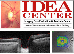
The UC San Diego’s Imaging Data Evaluation and Analysis (IDEA) Center offers a wide variety of clinical research support services from image processing and analysis to study design and protocol development support. Our highly skilled researchers and staff are expertly trained in several types of image processing and analysis.
The IDEA Center is also comprised of the Optic Disc Reading Center (ODRC), which offers interpretation and analysis of fundus photography, including optic disc and retinal photographs, both digital and film.
What We Do
Our areas of expertise include, but are not limited to, the processing and quality review of Optical Coherence Tomography; Swept-Source OCT; and fundus photography. We work closely with manufacturers and engineers of our imaging equipment and software to implement innovative technologies, improve workflows, and promote consistency across participating centers.
Services
The IDEA Center offers a menu of services to meet clinical study-specific needs. Linda Zangwill, PhD and the IDEA Center staff will work with the study's sponsors to provide the optimum complement of services to ensure successful data transfer, management, and processing of images and photographs.
Protocol Development
• Assist developing imaging and photography protocols
• Develop and maintain data collection forms and study documents
• Provide study-specific manual of procedures
• Provide protocols and training for electronic transfer of images and photographs
• Coordinate with instrument vendors to harmonize equipment and software across participating centers
Training and Certification
• Training of multi-center study personnel
• Certification of multi-center study personnel
Quality Assessment
• Review images and photographs for quality and adherence to protocol
• Provide quality control reports and actionable feedback to study centers to promote protocol adherence and data quality
• Request repeated testing or data corrections to ensure data completeness and accuracy
• Evaluate and pre-approve instrument hardware and software upgrades to ensure compatibility, performance, and study continuity
Data Handling
• FDA 21 CFR Part 11, HIPAA and IRB compliant data handling
• Study-specific image quality and protocol adherence assessment
• In-house IT /computer programming specialists available to create and maintain secure databases and provide support for all study needs
• Efficient electronic transfer of images and data
• Validate reading center processes to ensure accuracy, consistency, and regulatory compliance
Image Processing and Analysis
• Assess imaging scans for specific diagnoses and inclusion/exclusion criteria
• Perform expert correction of imaging scans, including segmentation, BMO positioning, and measurement centering
• Provide to sponsors study data and quality control results in user-friendly formats accompanied by comprehensive data dictionaries
• Provide progression analysis
• Provide statistical analysis
• Other Services
Contact Dr. Zangwill hgcreadingcenters@health.ucsd.edu to discuss services not listed above. A companion reading center, the VisFACT Center, is available to provide similar services for visual field tests.
Reading Center Studies
IDEA Center has provided reading center services for the following studies:
Sponsored by National Eye Institute
• Diagnostic Innovations in Glaucoma Study (DIGS): Glaucoma and High Myopia (2017-present)
• Diagnostic Innovations in Glaucoma Study (DIGS) (2003- present)
• Monitoring of Glaucoma Patients in Advanced Disease (2022-present)
• Multimodal Artificial Intelligence to Predict Glaucomatous Progression and Surgical Intervention (2022-2026)
• Diagnosis and Monitoring of Glaucoma with Optical Coherence Tomography Angiography (2018-2026)
African Descent and Glaucoma Evaluation Study (ADAGES)
• African Descent and Glaucoma Evaluation Study (ADAGES) IV: Alterations of the Lamina Cribrosa in Progression (2017-2022)
• African Descent and Glaucoma Evaluation Study (ADAGES) III: Contribution of Genotype to Glaucoma Phenotype in African Americans (2014-2019)
• African Descent and Glaucoma Evaluation Study (ADAGES II) (2009-2016)-AfricanDescent and Glaucoma Evaluation Study (ADAGES) (2003-2009)
The Ocular Hypertension Treatment Study (OHTS)
• The Ocular Hypertension Treatment Study: 20 Year Follow-up (2015-2019)
• Ocular Hypertension Treatment Study (OHTS) and European Glaucoma Prevention Study (EGPS): Glaucoma Detection using Confocal Scanning Laser Ophthalmoscopy (2008-2011)
• Ocular Hypertension Treatment Study Ancillary Investigation: Confocal Scanning Laser Ophthalmoscopy of the Optic Disc (1995-2010)
• Retinal Nerve Fiber Layer Thickness, Optic Nerve and Choroidal Thickness Measurements in the COMET Cohort of Young Myopic Adults Correction of Myopia Evaluation Trial (2009-2016)
Sponsored by Heidelberg Engineering, Inc
Myopia OCT Phenotyping (GMOPC): 2021-present
• Cohort 1: Multi-center study of optic nerve head, peripapillary, and posterior pole structure measured with the Heidelberg Spectralis OCT in healthy highly myopic eyes.
• Cohort 2: Multi-center study of optic nerve head, peripapillary, and posterior pole structure measured with the Heidelberg Spectralis OCT in highly myopic eyes with glaucoma.
• Cohort 3: Multi-center study of optic nerve head, peripapillary, and posterior pole structure measured with the Heidelberg Spectralis OCT within non-highly myopic eyes with early glaucoma.
• The Heidelberg Engineering ANTERION Anterior Segment Cornea and IOL Precision and Agreement Study (2020-2022)
• OCT Imaging Reading Center for FDA regulated study sponsored by Heidelberg Engineering Inc., “The Heidelberg Engineering ANTERION Imaging Agreement Study” (2019-2021)
Sponsored by Allergan Pharmaceuticals, Inc
• A 48-Month, Multi-Center, Randomized, Double-Masked Placebo-Controlled, Clinical Study to Evaluate the Effectiveness and Safety of Oral Memantine in Daily Doses of 20 MG and 10 MG in Patients with Chronic Open-Angle Glaucoma at Risk for Glaucomatous Progression. Protocol Number 192944-004.(1999-2008)
• A 48-Month, Multi-Center, Randomized, Double-Masked Placebo-Controlled, Clinical Study to Evaluate the Effectiveness and Safety of Oral Memantine in Daily Doses of 20 MG and 10 MG in Patients with Chronic Open-Angle Glaucoma at Risk for Glaucomatous Progression. Protocol Number 192944-005. (1999-2008)
Sponsored by TOPCON Corporation
• Topcon DRI-OCT-1 Optical Coherence Tomography System for the Acquisition of Retinal Thickness Measurements and Ocular Images of the Posterior Chamber: Agreement and Precision Study (2013-2016)
• An age-stratified data collectionstudy in adult males and females, ages 18 and above to establish a normativedatabase using the 3D optical coherence Tomography 3D OCT-1000 Mark II (2009-2010)
Sponsored by Nidek Co., Ltd (2011-2012)
• Normative Data Collection Study of the Nidek Optical Coherence Tomography RS-3000 for the Measurements of Retinal and RNFL Thickness and Optic Disc Analysis (2012-2014)
• Comparative Study of the Nidek Optical Coherence Tomography RS-3000 and the RTVue OCT Predicate Device for the Measurements of Retinal and RNFL Thickness, Optic Disc Analysis, Pachymetry, Anterior Chamber Imaging and SLO Imaging. (2011-2012)
Reading Center Personnel
- • Linda Zangwill, PhD; Director
- • Keri Dirkes, MPH; Managing Director
- • Maria Hunsicker; Coordinator
- • Suzanne Vega, MPH; Coordinator
Scientific Advisory Board
Our board is composed of internationally recognized ophthalmologists and researchers including:
- • Christopher Girkin, MD, MSPH
- • Robert N. Weinreb, MD
- • Linda M. Zangwill, PhD
Information about diversity and inclusion is available through multiple venues. First, there are several websites available that provide information about diversity initiatives across the entire campus (diversity.ucsd.edu) and the (School of Medicine). Many departments have their own websites as well. Virtually all departments at UCSD have identified an Equity, Diversity, and Inclusion (EDI) ambassador, typically a faculty member who spearheads diversity-related initiatives in their areas/specialties and also collaborates with various entities across the medical school.
There are several active affinity organizations for UCSD medical students. These include:
The Association of Native American Medical Students (ANAMS)
The Asian Pacific American Medical Students Association (APAMSA)LGBTQ
Pharmacy and Medical Students (LGBTQ-PhaM)
The Latino Medical Student Association (LMSA)
Medical Students for Justice (MS4J)
The Student National Medical Association (SNMA)
UCSD also offers a curricular pathway called Program in Medical Education – Health Equity (PRIME - HEq), which part of a system-wide effort at the University of California to train physicians better able to meet the needs of the diverse Californian population who are traditionally underserved by the medical system. PRIME-HEq faculty work with students to identify populations or communities at risk for health disparities. Students will then receive exposure, training, and the opportunity to work with the identified group to further their passion in the area and provide knowledge and skills to better equip the students to improve health equity for the group.
- UC Campus Diversity Efforts
- UCSD Office for Equity, Diversity, and Inclusion
- Center for Faculty Diversity and Inclusion
- UCSD School of Medicine Office of Diversity and Community Partnerships
- UCSD Student National Medical Association
- UCSD Black Resource Center
- UCSD Latino Medical Student Association
- UCSD Women’s Center
- UCSD Hispanic Center of Excellence
- UCSD LGBT Resource Center